
Structure dun muscle de copépode après un double stress thermique et ionique. Ibrahim A, Souissi A, Leray A, Héliot L, Souissi S, Vandenbunder B
Soumis le 30 Jul 2015 par Bernard Vandenbunder
Mot(s)-clé(s) : tissue, biomechanics
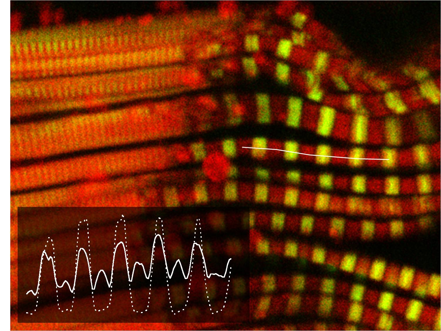
Functional anatomy of the muscle of a swimming champion, Pseudodiaptomus marinus copepod, following a mixed temperature and haline jump. Overlaid images of myofibrils showing flavin two-photon autofluorescence bands located between (red) and overlapping (yellow/green) myosin sarcomeric A bands visualized by their Second harmonic Generation signal. The insert displays the TPEF (solid lines) and SHG (dotted lines) signal intensity profiles along the line drawn through a myofibril. Note that P. marinus can reach 130 µm/s, a velocity equaling 100 body lengths s-1 during escape responses.