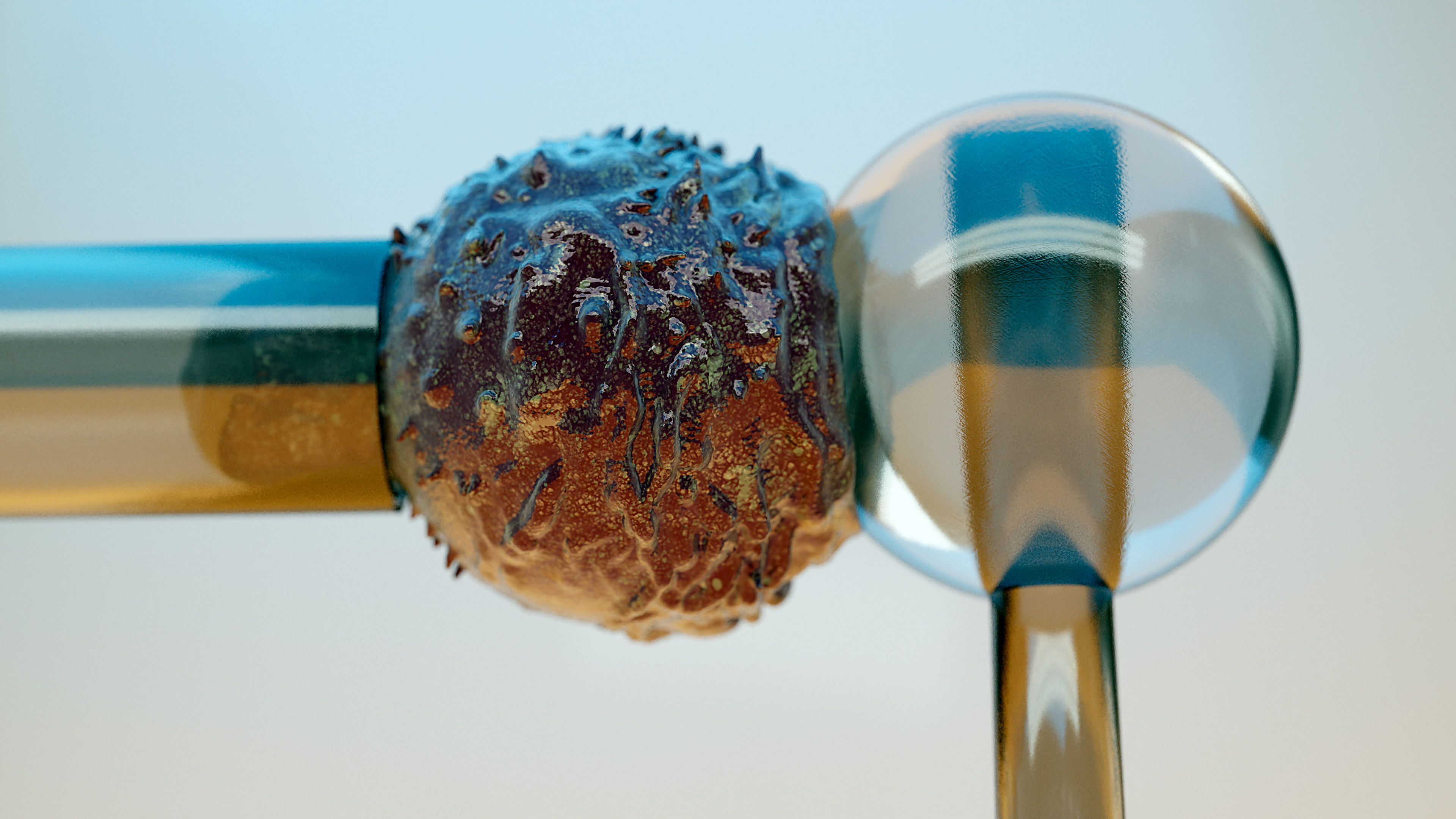
Micropipette force probe to quantify single-cell force generation: application to T-cell activation
A. Sawicka, A. Babataheri, S. Dogniaux, A. I. Barakat, D. Gonzalez-Rodriguez, C. Hivroz, and J. Husson
Molecular Biology of the Cell 28, 3229-3239 (2017) [Accès à la revue]
In response to engagement of surface molecules, cells generate active forces that regulate many cellular processes. Developing tools that permit gathering mechanical and morphological information on these forces is of the utmost importance. Here we describe a new technique, the Micropipette Force Probe, that uses a micropipette as a flexible cantilever that can aspirate at its tip a bead that is coated with molecules of interest and is brought in contact with the cell. This technique simultaneously allows tracking the resulting changes in cell morphology and mechanics as well as measuring the forces generated by the cell. To illustrate the power of this technique, we applied it to the study of human primary T lymphocytes (T cells). It allowed the fine monitoring of pushing and pulling forces generated by T cells in response to various activating antibodies and bending stiffness of the micropipette. We further dissected the sequence of mechanical and morphological events occurring during T cell activation to model force generation and to reveal heterogeneity in the cell population studied. We also report the first measurement of the changes in Young’s modulus of T cells during their activation, showing that T cells stiffen within the first minutes of the activation process.
Abstract: In response to engagement of surface molecules, cells generate active forces that regulate many cellular processes. Developing tools that permit gathering mechanical and morphological information on these forces is of the utmost importance. Here we describe a new technique, the Micropipette Force Probe, that uses a micropipette as a flexible cantilever that can aspirate at its tip a bead that is coated with molecules of interest and is brought in contact with the cell. This technique simultaneously allows tracking the resulting changes in cell morphology and mechanics as well as measuring the forces generated by the cell. To illustrate the power of this technique, we applied it to the study of human primary T lymphocytes (T cells). It allowed the fine monitoring of pushing and pulling forces generated by T cells in response to various activating antibodies and bending stiffness of the micropipette. We further dissected the sequence of mechanical and morphological events occurring during T cell activation to model force generation and to reveal heterogeneity in the cell population studied. We also report the first measurement of the changes in Young’s modulus of T cells during their activation, showing that T cells stiffen within the first minutes of the activation process.
Thème : Thème 2007-2010 : Mécanique cellulaire
Equipe : Génie Cellulaire et Cardiovasculaire (LadHyX)
